BloodWorX AI Live Blood Analysis Software
BloodWorX AI Live Blood Analysis Software
See What Your Blood Reveals. In minutes, Not Hours.
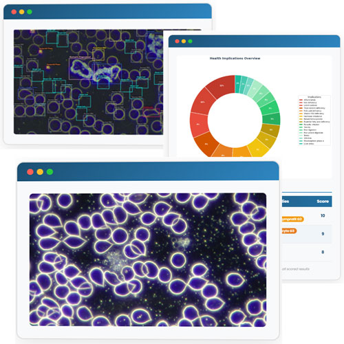
BloodWorX AI analyses live blood under darkfield microscopy, automatically detecting over 50 cell anomalies, generating professional reports with health implications, personalised action plans, and annotated sample images — all powered by advanced artificial intelligence.
Trained on 1M+ cells • 50+ anomaly types • Reports in under 3 minutes • English & Spanish
Educational & Supportive Tool — Not a Diagnostic Device
BloodWorX AI is designed to support trained practitioners with darkfield microscopy analysis. It does not provide medical diagnoses, treatment prescriptions, or replace the judgment of a qualified healthcare professional. All AI-generated findings are for educational and informational purposes only and must be interpreted by a trained practitioner.
Imagine this: your client sits across from you. A single drop of blood goes under the microscope.
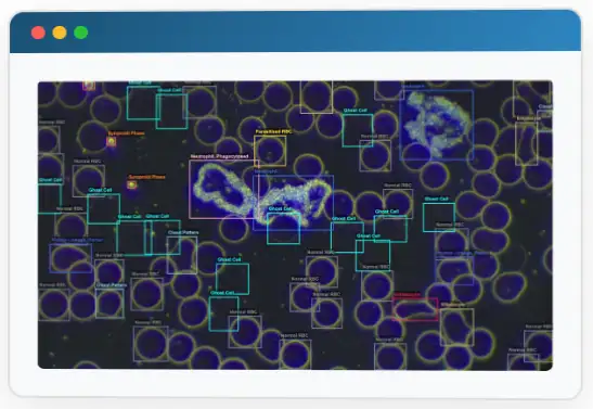
Within minutes, your screen lights up — every cell automatically detected, labelled, and graded. Ghost cells, elliptocytes, cloud patterns, protein linkages — all identified with confidence scores. No guessing. No searching through reference books.
Before your client finishes their glass of water, you hand them a beautifully formatted report: ranked health implications, a personalised action plan with dietary changes, supplement dosages, and lifestyle recommendations — plus the exact lab tests they should ask their doctor about.
That's not the future. That's BloodWorX AI right now. Here's exactly how it works, step by step — with real examples from an actual report.
The Journey — From Blood Drop to Full Report
A Drop of Blood, a World of Insight
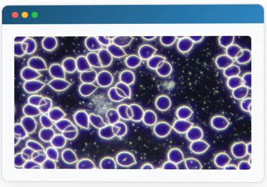
It starts with a simple finger prick. The fresh blood goes onto a slide, under your darkfield microscope — alive, unstained, in its natural state. You capture the image and upload it to BloodWorX AI. That's all you need to do.
- Works with any darkfield microscope setup
- Upload JPG, PNG, or TIFF — no special format needed
- Analyse multiple images per session
The AI Sees What the Naked Eye Misses
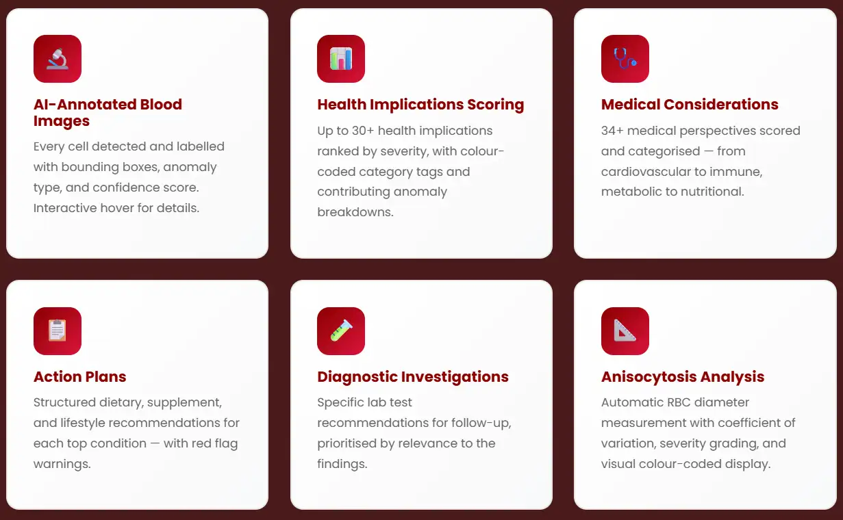
In minutes, our deep learning model (AI Model, trained on 1M+ cells) scans every corner of your image. Each cell gets a bounding box, a classification label, and a confidence score. Anomalies are graded by severity — from G1 (mild) to G5 (critical). Nothing escapes detection.
- Automatic bounding box annotations on every cell
- Confidence percentage for each detection
- Severity grading G1 through G5
- Cell-by-cell breakdown per uploaded image
From Cells to meaningful Meaning
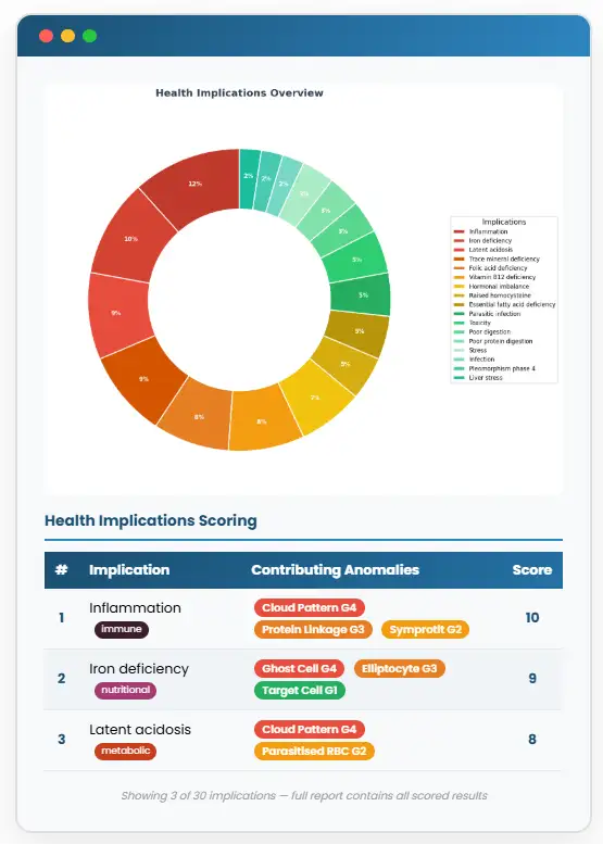
Here's where the magic happens. The AI doesn't just find anomalies — it connects the dots. Each detected cell is cross-referenced against a knowledge base. Inflammation? Iron deficiency? Latent acidosis? The system scores each health implication based on which anomalies contribute and how severe they are, then ranks them from most to least urgent.
- Up to 30+ health implications scored automatically
- Categorised: immune, nutritional, metabolic, and more
- Visual donut charts for at-a-glance overview
- Ranked tables showing exactly which anomalies drove each score
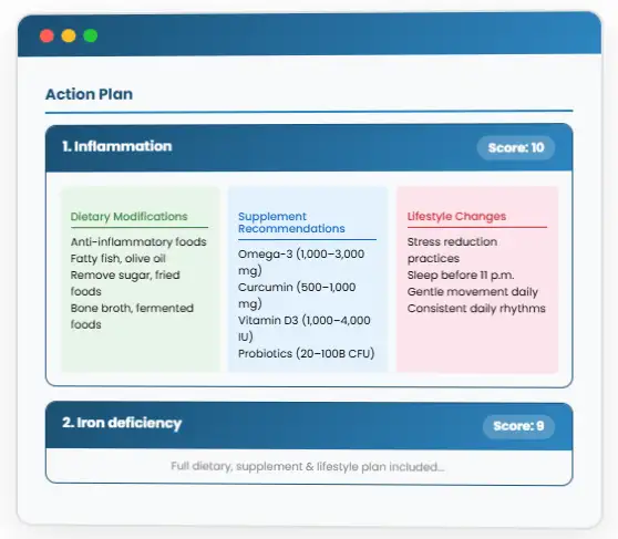
Your Client's Personal Roadmap to Health
This is what your clients will remember you for. For each top health implication, BloodWorX AI generates a detailed, three-pillar action plan: what to eat (and what to avoid), which supplements to take (with exact dosages), and which lifestyle habits to adopt. It even flags red-flag symptoms that need immediate medical referral.
- Priority-ordered by relevance
- Red flag warnings for emergency referral situations
- Exact supplement dosages and dietary protocols
- Print-ready — hand it directly to your client
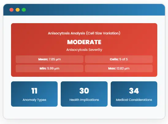
Measuring What Was Once Unmeasurable
Are the red blood cells all the same size? Or is there significant variation — a sign of nutritional deficiency or bone marrow stress? BloodWorX AI measures individual cell diameters in micrometres, calculates the coefficient of variation, and assigns a colour-coded severity grade. What used to be a rough visual estimate becomes precise, quantified data.
- Works with any darkfield microscope setup
- Mean RBC diameter (in μm)
- Minimum and maximum diameter range
- Coefficient of variation (CV) — the gold standard
- Colour-coded severity: Normal / Mild / Moderate / Severe
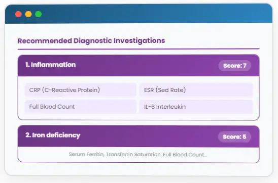
Bridging the Gap to Conventional Medicine
Your client wants to take the next step with their GP? BloodWorX AI recommends the exact lab tests to request — CRP, ferritin, full blood count, and more — prioritised by relevance to the darkfield findings. You're not just analysing blood; you're giving your client a bridge between holistic and conventional care.
- Condition-specific test panels (e.g., CRP, ESR, IL-6)
- Priority scoring per investigation
- Evidence-based lab test recommendations
- Empowers clients to have informed conversations with their GP
The whole process? Under 3 Minutes.
Your client is still sitting across from you. But now they're holding a multi-page report — with annotated images of their own blood cells, a ranked list of health implications, a personalised action plan, and the exact tests to discuss with their doctor.
They didn't wait days. They didn't need a lab. They got answers — clear, visual, actionable — while the blood was still alive on the slide.
That's the experience your clients will talk about. That's what makes them come back — and refer others.
What's in Every BloodWorX AI Report
BloodWorX AI vs. Manual Analysis
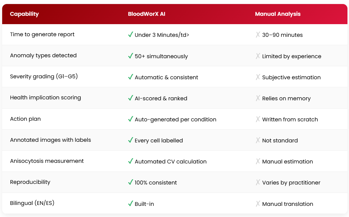
All AI-generated outputs are educational support tools. They do not constitute medical diagnosis and must be interpreted by a qualified practitioner
50+ Detectable Cell Anomalies
The AI automatically identifies and classifies all of the following cell anomalies from a single darkfield microscopy image.
Red Blood Cells (Erythrocytes)
- Normal
- Acanthocyte
- Echinocyte
- Ovalocyte
- Spherocyte
- Stomatocyte
- Target Cell
- Schistocyte
- Rouleaux
- Ring RBC
- Colloid Bodies
- Ghost Cell
- Elliptocyte
Aggregations & Formations
- Erythrocyte Aggregation
- Platelet Aggregation
- Platelet Agg. Fermentation
- Early Platelet Agg. Ferm.
- Cloud Pattern
- Complete Protein Linkage
- RBC Protein Linkage
- Pre-Rouleaux
White Blood Cells (Leukocytes)
- Basophil
- Eosinophil
- Lymphocyte
- Monocyte
- Neutrophil
- Band Neutrophil
- Degenerated Neutrophil
- Cohesive Neutrophil
- Neutrophil, Phagocytosed
- Ghost Cell
- Keratocyte
Microorganisms & Parasites
- Bacterial Form
- Symprotit Phase
- Parasitised RBC
- Chondrit
Crystals, Structures & Other
- Cholesterol Crystal
- Fat Cluster
- Fat Microdroplet
- Fibrin
- Fibrin Nest
- Metal
- Protoplast
- Pteroharpen
- Platelets
- Poikilocytes
- Membrane Protrusion
- Fermentation
Stop Spending Hours on Reports. Start Impressing Clients in minutes
Practitioners worldwide are already using BloodWorX AI to deliver faster, more accurate, and more comprehensive darkfield microscopy reports. The AI does the heavy lifting — you focus on what matters: your client.
Trained on 1M+ cells • 50+ anomaly types • Reports in under 3 minutes • English & Spanish
Important: BloodWorX AI is an educational and supportive tool designed to complement — not replace — professional judgement. Reports do not constitute medical diagnosis, treatment advice, or a substitute for consultation with a qualified healthcare professional. All findings should be interpreted by a trained practitioner and are intended for informational purposes only.
Related products
-
Sale!
BBF Real Rife Therapy System
Original price was: USD$5,170.00.USD$4,653.00Current price is: USD$4,653.00. -
Sale!

BBF 12-Week Live Blood Online Training Course
Original price was: USD$995.00.USD$895.50Current price is: USD$895.50. -
Sale!

Live Blood Analysis: BBF-HDMI-LED Package
Original price was: USD$11,301.00.USD$10,170.90Current price is: USD$10,170.90.